Scientists have developed a faster method to create highly detailed 3D models of ants by combining advanced X-ray imaging, robotics and artificial intelligence, enabling researchers to digitally reconstruct hundreds of species in a fraction of the usual time.
The study, published on March 5 in the journal Nature Methods, was led by researchers including Evan Economo of the University of Maryland and Thomas van de Kamp of the Karlsruhe Institute of Technology (KIT) in Germany.
For more than a decade, Economo’s laboratory has relied on micro-CT scanners to examine insect morphology—the study of their physical structures. Although the technology produces extremely detailed 3D images, scanning a single specimen can take up to 10 hours.
To overcome this limitation, the team developed a high-throughput system integrating a synchrotron particle accelerator, X-ray imaging, robotics and AI. The approach allowed scientists to rapidly scan thousands of ant specimens and convert the data into interactive 3D models.
The project, called Antscan, produced digital reconstructions of around 800 ant species. Researchers said completing such a task using conventional CT scanners would have required roughly six years of continuous work.
Instead, using facilities at KIT, the team scanned about 2,000 specimens in just one week.
The ants, preserved in ethanol, were collected from museums, partner institutions and private specialists worldwide. The specimens were then transported to KIT, where a powerful synchrotron beam generated intense X-rays capable of imaging multiple samples rapidly.
A robotic system handled the specimens during scanning, rotating each sample and replacing it with another every 30 seconds. The process produced stacks of two-dimensional images that were later combined into detailed 3D reconstructions.
However, many of the ants initially appeared in distorted positions after scanning. To address this, computer science students developed AI tools that automatically corrected the insects’ posture, producing lifelike digital models resembling ants in their natural positions.
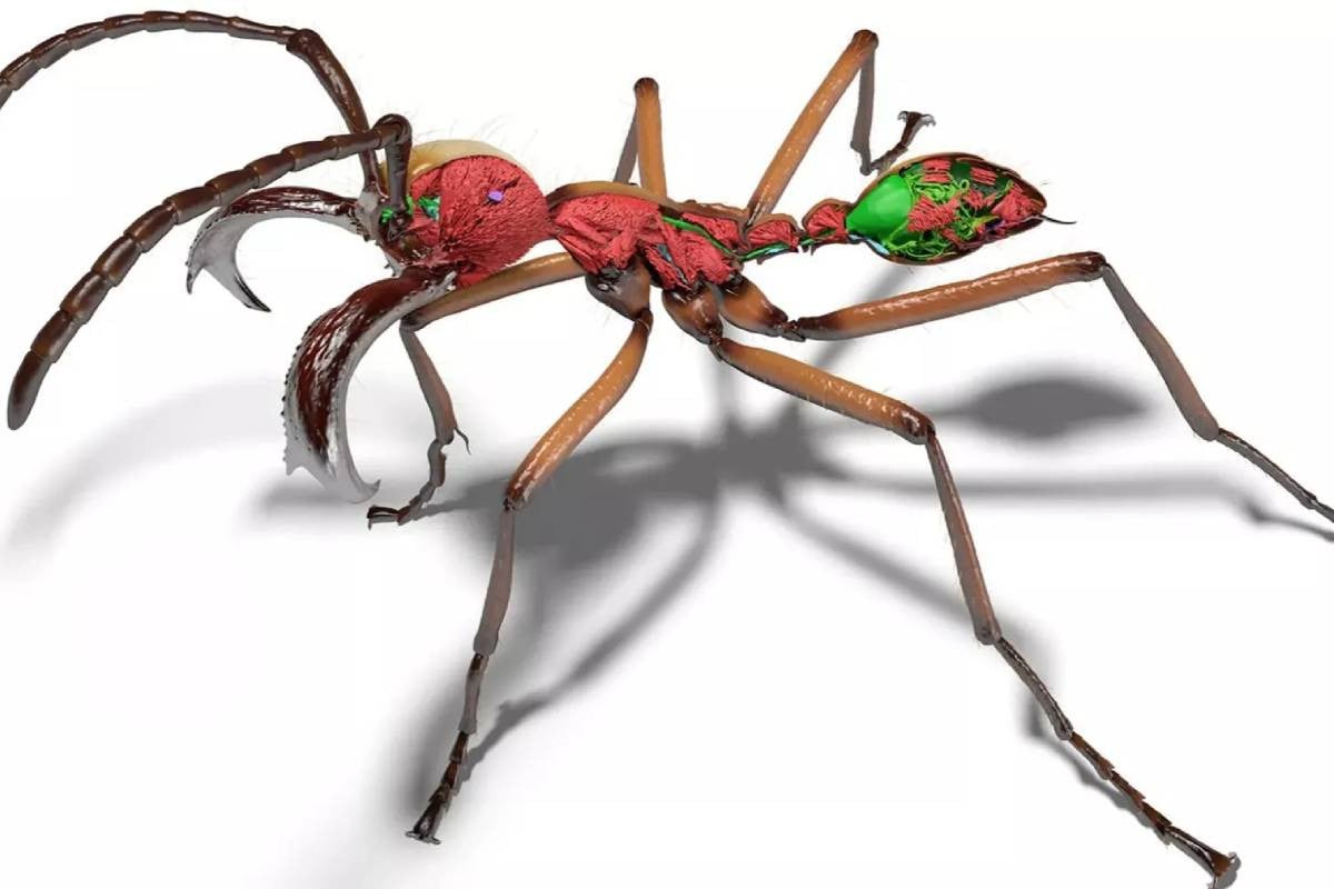
The resulting models reveal internal structures—including muscles, nervous systems, digestive organs and stingers—at micrometer-level resolution. Scientists say the digital ants can also be animated or placed in virtual reality environments for research, education and visual media production.
The Antscan database has already supported new scientific work. In a separate study published in Science Advances in December 2025, researchers used the data to examine how ant colonies balance worker size and physical strength.
By analysing more than 500 species, the team found that colonies investing less in thick exoskeleton armour often maintain larger numbers of workers. The findings suggest that lower investment in protective cuticle may allow colonies to expand and diversify more successfully.
Researchers say the detailed 3D models allow precise measurement of structures such as cuticle volume—something that was previously difficult to calculate.
Scientists believe the growing Antscan archive could eventually serve as a digital library of biodiversity. The scans may also help train machine-learning systems to automatically identify ant species during field research.
The team plans to expand the database by scanning more specimens and applying similar AI-based techniques to other biological datasets, potentially opening new avenues for studying the diversity of life on Earth.
Source: Science Daily
Bd-pratidin English/ Jisan