Skin cancer cases are rising in South Asia, despite generally lower rates due to higher melanin levels. Increased UV exposure and late detection are key contributing factors.
In an interview with the New York Post, board-certified dermatologist Dr. Asha Patel Shah shared the key warning signs of various types of skin cancer and pointed out skin changes that shouldn't be ignored. She also offered a step-by-step guide for self-examination, including the ‘ABCDEs’ to watch for. Since skin cancer is among the most preventable cancers, following Dr. Patel Shah’s advice can help protect you and your family.
What are the types of skin cancer?
There are three main types of skin cancer — basal cell carcinoma (BCC), squamous cell carcinoma (SCC) and melanoma.
BCC is the most common skin cancer.
It develops from basal cells in the deepest layer of the skin and looks like a small, pearly or flesh-colored bump or a reddish-pink scaly patch.
SCC stems from squamous cells in the upper layer of the skin.
It resembles rough, red patches or firm, pink bumps.
Melanoma is the most dangerous form of skin cancer.
“Skin cancer can truly mimic benign skin lesions, conditions or symptoms,” said Patel Shah, head of medical affairs North America, skin health & beauty at the consumer health company Kenvue.
Self-examination guide
“I would suggest starting with a baseline skin self-exam done at home (and with assistance from a mirror and/or partner) to really learn the patterns of your own skin,” Dr. Asha Patel Shah said to New York Post, “That way, when something changes, you can compare it to your own known baseline.” She mentions that it is important not to overlook any areas of the skin during a self check, which includes areas like the scalp, genitals, palms, underneath the fingernails and breasts, and between your fingers.
It is tricky to identify skin cancers from innocent lesions, hence its useful to keep in mind the ‘ABCDEs’ of skin cancer while performing a self exam, as stated by the dermatologist:
- Asymmetry - One half of the lesion looks different from the other.
- Border - The edges of the lesion or mole are poorly defined or irregular.
- Colour - Colours of the moles are irregular, or there are multiple colours in a single mole.
- Diameter - The width is about the size of a pencil eraser, larger than ¼ inch.
- Evolving - The shape, size and colour of the lesions change over time.
If you notice something abnormal or akin to the above descriptions, send a photo to your doctor immediately. The dermatologist suggests to carry out self examinations every month, especially for those who have previous or family history of skin cancer, a history of abnormal moles or immunosuppressed patients. A yearly dermatologist visit should be mandatory for those more at risk.
Note these abnormalities
Patel Shah wants you to keep an eye on these skin changes.
- A growth, sore or lesion that does not heal, keeps reopening after healing or bleeds/scabs and never heals
- A rough, dry patch resistant to moisturizers, especially if it is in a sun-exposed area of the body like the face, forearms or chest
- A rapidly growing new lump, bump or growth that may be painful or sore
- Persistent wart-like growths that may be painful or sore
- Pigmented lesions that have signs of asymmetry, irregular borders or color changes
- Pigmented lesions on “special sites” like the palms or soles
- Dark, uneven or wide streaks in the fingernail plates
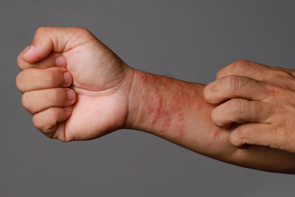
- Anything on the skin that is constantly itchy, sore, tender, painful and/or bleeding
- Cases of widespread, treatment-resistant skin conditions resembling eczema that could indicate a skin cancer that is not related to UV exposure, though this is rare.
Source: New York Post
Bd-pratidin English/ ANI